

Which Of The Following Is True About The Cytoplasm Of An Animal Oocyte
| Oocyte | |
|---|---|
| Identifiers | |
| MeSH | D009865 |
| FMA | 18644 |
| Anatomical terminology [edit on Wikidata] | |
An oocyte (, ), oöcyte, or ovocyte is a female gametocyte or germ cell involved in reproduction. In other words, it is an immature ovum, or egg jail cell. An oocyte is produced in a female person fetus in the ovary during female gametogenesis. The female germ cells produce a primordial germ cell (PGC), which then undergoes mitosis, forming oogonia. During oogenesis, the oogonia become principal oocytes. An oocyte is a course of genetic material that tin be nerveless for cryoconservation.
Formation [edit]
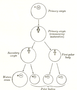
Diagram showing the reduction in number of the chromosomes in the process of maturation of the ovum; the process is known equally meiosis.
The formation of an oocyte is chosen oocytogenesis, which is a role of oogenesis.[i] Oogenesis results in the formation of both principal oocytes during fetal period, and of secondary oocytes after it as part of ovulation.
| Prison cell type | ploidy/chromosomes | chromatids | Process | Time of completion |
| Oogonium | diploid/46(2N) | 2C | Oocytogenesis (mitosis) | third trimester |
| primary Oocyte | diploid/46(2N) | 4C | Ootidogenesis (meiosis I) (Folliculogenesis) | Dictyate in prophase I for up to 50 years |
| secondary Oocyte | haploid/23(1N) | 2C | Ootidogenesis (meiosis II) | Halted in metaphase Ii until fertilization |
| Ootid | haploid/23(1N) | 1C | Ootidogenesis (meiosis II) | Minutes after fertilization |
| Ovum | haploid/23(1N) | 1C |
Characteristics [edit]
Cytoplasm [edit]
Oocytes are rich in cytoplasm, which contains yolk granules to nourish the cell early in evolution.
Nucleus [edit]
During the chief oocyte phase of oogenesis, the nucleus is chosen a germinal vesicle.[2]
The only normal human type of secondary oocyte has the 23rd (sex) chromosome every bit 23,10 (female-determining), whereas sperm tin accept 23,X (female-determining) or 23,Y (male-determining).
Nest [edit]
The space within an ovum or young ovum is located is the cell-nest.[3]
Cumulus-Oocyte Complex [edit]
The cumulus-oocyte complex contains layers of tightly packed cumulus cells surrounding the oocyte in the Graafian follicle. The oocyte is arrested in Meiosis Two at the stage of metaphase Ii and is considered a secondary oocyte. Before ovulation, the cumulus complex goes through a structural change known as cumulus expansion. The granulosa cells transform from tightly compacted to an expanded mucoid matrix. Many studies show that cumulus expansion is critical for the maturation of the oocyte because the cumulus complex is the oocyte's direct advice with the developing follicle surroundings. It also plays a pregnant role in fertilization, though the mechanisms are not entirely known and are species specific.[4] [v] [6]
Maternal contributions [edit]

Because the fate of an oocyte is to become fertilized and ultimately grow into a fully operation organism, it must be ready to regulate multiple cellular and developmental processes. The oocyte, a large and complex cell, must be supplied with numerous molecules that will straight the growth of the embryo and control cellular activities. As the oocyte is a product of female gametogenesis, the maternal contribution to the oocyte and consequently the newly fertilized egg, is enormous. There are many types of molecules that are maternally supplied to the oocyte, which will direct various activities inside the growing zygote.
Avoidance of impairment to germ-line Deoxyribonucleic acid [edit]
The Dna of a prison cell is vulnerable to the dissentious effect of oxidative gratis radicals produced equally byproducts of cellular metabolism. Deoxyribonucleic acid harm occurring in oocytes, if not repaired, can be lethal and result in reduced fecundity and loss of potential progeny. Oocytes are essentially larger than the boilerplate somatic jail cell, and thus considerable metabolic activeness is necessary for their provisioning. If this metabolic activeness were carried out by the oocyte's own metabolic machinery, the oocyte genome would be exposed to the reactive oxidative by-products generated. Thus it appears that a process evolved to avoid this vulnerability of germ line DNA. It was proposed that, in gild to avoid damage to the DNA genome of the oocytes, the metabolism contributing to the synthesis of much of the oocyte's constituents was shifted to other maternal cells that so transferred these constituents to oocytes.[seven] [8] Thus, oocytes of many organisms are protected from oxidative DNA damage while storing up a big mass of substances to nurture the zygote in its initial embryonic growth.
mRNAs and proteins [edit]
During the growth of the oocyte, a diverseness of maternally transcribed messenger RNAs, or mRNAs, are supplied by maternal cells. These mRNAs can be stored in mRNP (message ribonucleoprotein) complexes and be translated at specific time points, they can exist localized within a specific region of the cytoplasm, or they can be homogeneously dispersed within the cytoplasm of the unabridged oocyte.[9] Maternally loaded proteins can also be localized or ubiquitous throughout the cytoplasm. The translated products of the mRNAs and the loaded proteins accept multiple functions; from regulation of cellular "house-keeping" such as prison cell cycle progression and cellular metabolism, to regulation of developmental processes such every bit fertilization, activation of zygotic transcription, and germination of body axes.[9] Beneath are some examples of maternally inherited mRNAs and proteins constitute in the oocytes of the African clawed frog.
| Name | Type of maternal molecule | Localization | Function |
|---|---|---|---|
| VegT[10] | mRNA | Vegetal hemisphere | Transcription factor |
| Vg1[11] | mRNA | Vegetal hemisphere | Transcription cistron |
| XXBP-ane[12] | mRNA | Not known | Transcription factor |
| CREB[xiii] | Protein | Ubiquitous | Transcription factor |
| FoxH1[14] | mRNA | Ubiquitous | Transcription factor |
| p53[fifteen] | Poly peptide | Ubiquitous | Transcription Gene |
| Lef/Tcf[16] | mRNA | Ubiquitous | Transcription factor |
| FGF2[17] | Protein | Nucleus | Not known |
| FGF2, 4, 9 FGFR1[16] | mRNA | Not known | FGF signaling |
| Ectodermin[xviii] | Poly peptide | Animal hemisphere | Ubiquitin ligase |
| PACE4[19] | mRNA | Vegetal hemisphere | Proprotein convertase |
| Coco[20] | Poly peptide | Not known | BMP inhibitor |
| Twisted gastrulation[sixteen] | Protein | Non known | BMP/Chordin bounden protein |
| fatvg[21] | mRNA | Vegetal hemisphere | Germ cell formation and cortical rotation |

Mitochondria [edit]
The oocyte receives mitochondria from maternal cells, which volition go along to control embryonic metabolism and apoptotic events.[9] The partitioning of mitochondria is carried out by a organization of microtubules that will localize mitochondria throughout the oocyte. In certain organisms, such every bit mammals, paternal mitochondria brought to the oocyte past the spermatozoon are degraded through the attachment of ubiquitinated proteins. The destruction of paternal mitochondria ensures the strictly maternal inheritance of mitochondria and mitochondrial Dna or mtDNA.[9]
Nucleolus [edit]
In mammals, the nucleolus of the oocyte is derived solely from maternal cells.[22] The nucleolus, a structure found within the nucleus, is the location where rRNA is transcribed and assembled into ribosomes. While the nucleolus is dense and inactive in a mature oocyte, it is required for proper development of the embryo.[22]
Ribosomes [edit]
Maternal cells also synthesize and contribute a store of ribosomes that are required for the translation of proteins before the zygotic genome is activated. In mammalian oocytes, maternally derived ribosomes and some mRNAs are stored in a structure called cytoplasmic lattices. These cytoplasmic lattices, a network of fibrils, poly peptide, and RNAs, have been observed to increase in density as the number of ribosomes subtract within a growing oocyte.[23]
Prophase I arrest [edit]
Female mammals and birds are born possessing all the oocytes needed for hereafter ovulations, and these oocytes are arrested at the prophase I phase of meiosis.[24] In humans, as an case, oocytes are formed between three and iv months of gestation within the fetus and are therefor nowadays at birth. During this prophase I arrested phase (dictyate), which may final for many years, four copies of the genome are nowadays in the oocytes. The abort of ooctyes at the four genome copy stage appears to provide the informational back-up needed to repair damage in the Deoxyribonucleic acid of the germline.[24] The repair process used likely involves homologous recombinational repair.[24] [25] [26] Prophase arrested oocytes take a loftier capability for efficient repair of Dna amercement.[25] DNA repair capability appears to exist a central quality command mechanism in the female person germ line and a critical determinant of fertility.[25]
Paternal contributions [edit]
The spermatozoon that fertilizes an oocyte will contribute its pronucleus, the other half of the zygotic genome. In some species, the spermatozoon will also contribute a centriole, which will aid brand upward the zygotic centrosome required for the first sectionalisation. However, in some species, such as in the mouse, the unabridged centrosome is acquired maternally.[27] Currently nether investigation is the possibility of other cytoplasmic contributions fabricated to the embryo by the spermatozoon.
During fertilization, the sperm provides 3 essential parts to the oocyte: (1) a signalling or activating cistron, which causes the metabolically fallow oocyte to activate; (2) the haploid paternal genome; (3) the centrosome, which is responsible for maintaining the microtubule organization. Meet anatomy of sperm
Abnormalities [edit]
- Nondisjunction—a failure of proper homolog separation in meiosis I, or sister chromatid separation in meiosis II can lead to aneuploidy, in which the oocyte has the wrong number of chromosomes, for example 22,X or 24,X. This is the cause of atmospheric condition like Down syndrome and Edwards syndrome in humans. It is more than likely with avant-garde maternal age.
- Some oocytes have multiple nuclei, although it is idea they never mature.
See also [edit]
- Cortical granule
- Cryoconservation of fauna genetic resources
- Folliculogenesis
- Oocyte maturation inhibitor
- Polar body
- Symmetry breaking and cortical rotation
References [edit]
- ^ answers.com
- ^ "Germinal vesicle". Biology Articles, Tutorials & Dictionary Online. 2019-10-07. Retrieved 2022-04-09 .
- ^ Grier HJ, Uribe MC, Parenti LR (Apr 2007). "Germinal epithelium, folliculogenesis, and postovulatory follicles in ovaries of rainbow trout, Oncorhynchus mykiss (Walbaum, 1792) (Teleostei, protacanthopterygii, salmoniformes)". Journal of Morphology. 268 (iv): 293–310. doi:10.1002/jmor.10518. PMID 17309079. S2CID 23482731.
- ^ Yokoo M, Sato E (2004). "Cumulus-oocyte complex interactions during oocyte maturation". International Review of Cytology. 235: 251–91. doi:10.1016/S0074-7696(04)35006-0. ISBN978-0-12-364639-2. PMID 15219785.
- ^ Tanghe S, Van Soom A, Nauwynck H, Coryn M, de Kruif A (March 2002). "Minireview: Functions of the cumulus oophorus during oocyte maturation, ovulation, and fertilization". Molecular Reproduction and Development. 61 (3): 414–24. doi:10.1002/mrd.10102. PMID 11835587. S2CID 5728551.
- ^ Huang Z, Wells D (October 2010). "The human oocyte and cumulus cells relationship: new insights from the cumulus cell transcriptome". Molecular Human Reproduction. 16 (10): 715–25. doi:10.1093/molehr/gaq031. PMID 20435609.
- ^ Bernstein C (1993). "Sexual activity equally a response to oxidative DNA damage. Affiliate 10". In Halliwell B, Aruoma OI (eds.). Dna and Free Radicals. Dandy United kingdom: Ellis Horwood Limited. pp. 204–205. ISBN978-0-13-222035-iv.
- ^ Bernstein C (1998). "Sex as a response to oxidative DNA harm. Chapter iv". In Aruoma OI, Halliwell B (eds.). DNA and Costless Radicals: Techniques, Mechanisms & Applications. Saint Lucia and London: OICA International. pp. 112–113. ISBN976-8056169.
- ^ a b c d Mtango NR, Potireddy S, Latham KE (2008). "Oocyte quality and maternal control of development". International Review of Cell and Molecular Biology. 268: 223–90. doi:10.1016/S1937-6448(08)00807-one. PMID 18703408.
- ^ Zhang J, Rex ML (December 1996). "Xenopus VegT RNA is localized to the vegetal cortex during oogenesis and encodes a novel T-box transcription cistron involved in mesodermal patterning". Development. 122 (12): 4119–29. doi:10.1242/dev.122.12.4119. PMID 9012531.
- ^ Heasman J, Wessely O, Langland R, Craig EJ, Kessler DS (December 2001). "Vegetal localization of maternal mRNAs is disrupted by VegT depletion". Developmental Biology. 240 (2): 377–86. doi:10.1006/dbio.2001.0495. PMID 11784070.
- ^ Zhao H, Cao Y, Grunz H (May 2003). "Xenopus Ten-box bounden protein 1, a leucine zipper transcription gene, is involved in the BMP signaling pathway". Developmental Biology. 257 (ii): 278–91. doi:10.1016/s0012-1606(03)00069-1. PMID 12729558.
- ^ Sundaram N, Tao Q, Wylie C, Heasman J (September 2003). "The function of maternal CREB in early embryogenesis of Xenopus laevis". Developmental Biology. 261 (two): 337–52. doi:x.1016/s0012-1606(03)00303-eight. PMID 14499645.
- ^ Kofron M, Puck H, Standley H, Wylie C, Quondam R, Whitman Yard, Heasman J (Oct 2004). "New roles for FoxH1 in patterning the early embryo". Evolution. 131 (20): 5065–78. doi:x.1242/dev.01396. PMID 15459100.
- ^ Takebayashi-Suzuki One thousand, Funami J, Tokumori D, Saito A, Watabe T, Miyazono 1000, et al. (September 2003). "Coaction between the tumor suppressor p53 and TGF beta signaling shapes embryonic torso axes in Xenopus". Evolution. 130 (17): 3929–39. doi:x.1242/dev.00615. PMID 12874116.
- ^ a b c Heasman J (February 2006). "Maternal determinants of embryonic cell fate". Seminars in Cell & Developmental Biology. 17 (1): 93–8. doi:10.1016/j.semcdb.2005.eleven.005. PMID 16426874.
- ^ Song J, Slack JM (December 1994). "Spatial and temporal expression of basic fibroblast growth factor (FGF-2) mRNA and protein in early on Xenopus evolution". Mechanisms of Development. 48 (3): 141–51. doi:10.1016/0925-4773(94)90055-8. PMID 7893598. S2CID 20281053.
- ^ Dupont S, Zacchigna L, Cordenonsi 1000, Soligo South, Adorno G, Rugge Thousand, Piccolo S (April 2005). "Germ-layer specification and control of cell growth by Ectodermin, a Smad4 ubiquitin ligase". Cell. 121 (i): 87–99. doi:10.1016/j.cell.2005.01.033. PMID 15820681. S2CID 16628152.
- ^ Birsoy B, Berg 50, Williams PH, Smith JC, Wylie CC, Christian JL, Heasman J (February 2005). "XPACE4 is a localized pro-protein convertase required for mesoderm induction and the cleavage of specific TGFbeta proteins in Xenopus development". Development. 132 (3): 591–602. doi:10.1242/dev.01599. PMID 15634697.
- ^ Bong E, Muñoz-Sanjuán I, Altmann CR, Vonica A, Brivanlou AH (Apr 2003). "Cell fate specification and competence by Coco, a maternal BMP, TGFbeta and Wnt inhibitor". Development. 130 (7): 1381–9. doi:x.1242/dev.00344. PMID 12588853.
- ^ Chan AP, Kloc M, Larabell CA, LeGros M, Etkin LD (May 2007). "The maternally localized RNA fatvg is required for cortical rotation and germ prison cell formation". Mechanisms of Development. 124 (5): 350–63. doi:10.1016/j.mod.2007.02.001. PMC2435194. PMID 17376659.
- ^ a b Ogushi South, Palmieri C, Fulka H, Saitou M, Miyano T, Fulka J (February 2008). "The maternal nucleolus is essential for early embryonic development in mammals". Science. 319 (5863): 613–6. doi:10.1126/science.1151276. PMID 18239124. S2CID 7799743.
- ^ Yurttas P, Vitale AM, Fitzhenry RJ, Cohen-Gould L, Wu West, Gossen JA, Coonrod SA (August 2008). "Function for PADI6 and the cytoplasmic lattices in ribosomal storage in oocytes and translational control in the early mouse embryo". Evolution. 135 (15): 2627–36. doi:10.1242/dev.016329. PMC2708103. PMID 18599511.
- ^ a b c Mira A (September 1998). "Why is meiosis arrested?". Journal of Theoretical Biology. 194 (ii): 275–87. Bibcode:1998JThBi.194..275M. doi:10.1006/jtbi.1998.0761. PMID 9778439.
- ^ a b c Stringer JM, Winship A, Zerafa N, Wakefield One thousand, Hutt K (May 2020). "Oocytes can efficiently repair Dna double-strand breaks to restore genetic integrity and protect offspring wellness". Proceedings of the National Academy of Sciences of the United states of America. 117 (21): 11513–11522. doi:10.1073/pnas.2001124117. PMC7260990. PMID 32381741.
- ^ He, Da-Jian; Wang, Lin; Zhang, Zhi-Bi; Guo, Kun; Li, Jing-Zheng; He, Xie-Chao; Cui, Qing-Hua; Zheng, Ping (2018-eleven-18). "Maternal gene Ooep may participate in homologous recombination-mediated DNA double-strand intermission repair in mouse oocytes". Zoological Research. 39 (6): 387–395. doi:10.24272/j.issn.2095-8137.2018.067. PMC6085769. PMID 29955025.
- ^ Sutovsky P, Schatten Grand (2000). "Paternal contributions to the mammalian zygote: fertilization after sperm-egg fusion". International Review of Cytology. 195: 1–65. doi:x.1016/s0074-7696(08)62703-5. ISBN978-0-12-364599-nine. PMID 10603574.
Sources [edit]
- Purves WK, Orians GH, Sadava D, Heller HC (2004). Life: The Science of Biology (7th ed.). Freeman, W. H. & Company. pp. 823–824. ISBN978-0-7167-9856-9.
External links [edit]
- Micrograph of a principal oocyte and follicle of a monkey
Source: https://en.wikipedia.org/wiki/Oocyte
Posted by: hambywherfust.blogspot.com
0 Response to "Which Of The Following Is True About The Cytoplasm Of An Animal Oocyte"
Post a Comment